Home
Uncategories
Anterior Shoulder Tendon Anatomy - Alternatives To Rotator Cuff Tear Surgery The Evidence For Non Surgical Options Caring Medical Florida
Anterior Shoulder Tendon Anatomy - Alternatives To Rotator Cuff Tear Surgery The Evidence For Non Surgical Options Caring Medical Florida
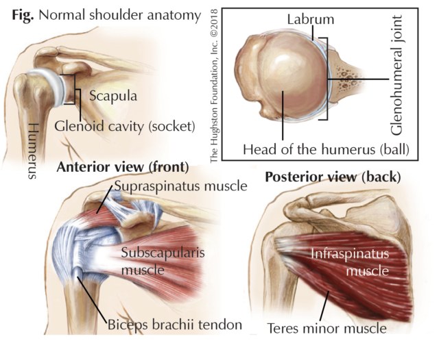
Anterior Shoulder Tendon Anatomy - Alternatives To Rotator Cuff Tear Surgery The Evidence For Non Surgical Options Caring Medical Florida. The shoulder joint, also known as the glenohumeral joint, is a ball and socket joint with the most extensive range of motion in the human body. The cervical spine and the biceps tendon. 11 photos of the shoulder muscles tendons anatomy. (the biceps is the muscle that weightlifters are always showing off). Shoulder tendon anatomy (page 1).
They connect your upper arm bone to your shoulder blade. The cervical spine and the biceps tendon. It is the part of the shoulder that borders the chest muscles. The anatomy of the lhbt and its corresponding structures has been extremely well researched. Its main function is shoulder flexion, which is characterized by raising your upper arms up to the front and overhead.
I Ve Jacked Up My Shoulder What Did I Damage And What Do I Do Now Breaking Muscle from cdn3.omidoo.com The rotator cuff tendons can be irritated or damaged. The term anterior shoulder instability refers to a shoulder in which soft tissue or bony insult allows the humeral head to sublux or dislocate from the glenoid fossa. The shoulder joint (glenohumeral joint) is a ball and socket joint between the scapula and the humerus. Axillary nerve (c5,6) from posterior cord of brachial plexus. A tendon is a structure that connects muscle to bone, and the biceps are connected by tendons at both the elbow and shoulder joints. Also in its upper part, the anterior portion of the subacromial subdeltoid bursa can be seen deep to the deltoid muscle and anterior to the biceps sheath. The scapula, clavicle and humerus are the bones of the shoulder. The upper portion of the bicep also has a tendon that attaches it to the bones within the shoulder.
In this condition the rotator cuff is unable to support the glenohumeral joint thereby causing pain in the biceps and the shoulder.
The shoulder is extremely mobile and made up of several joints that work together. On the anterior side of the shoulder, the coracobrachialis, serratus anterior, pectoralis major, and pectoralis minor muscles work as a group to flex and adduct the scapula and humerus anteriorly toward the sternum. The axillary nerve runs along the surgical neck of the humerus. Shoulder anatomy for ultrasound evaluation. A tendon is a structure that connects muscle to bone, and the biceps are connected by tendons at both the elbow and shoulder joints. The rotator cuff is a collection of muscles and tendons that surround the shoulder, giving it support and allowing a wide range of motion. A serious complication of shoulder dislocation is axillary nerve injury. The shoulder is made out of a ball and socket joint created by the scapula, humerus, and the muscles, ligaments, and tendons that support those bones. All of them are supplied by the respective branches of the brachial plexus. The shoulder joint is located between the glenoid fossa of the scapula and the humerus. The cervical spine and the biceps tendon. It works to allow a lot of range of motion in forward flexion (arms in front. The anterior deltoid is located on the front of your shoulder.
It is an injury to the glenohumeral joint (ghj) where the humerus is displaced from its normal position in the center of the glenoid fossa and the joint surfaces no longer touch each other. The anatomy of the lhbt and its corresponding structures has been extremely well researched. Its main function is shoulder flexion, which is characterized by raising your upper arms up to the front and overhead. This usually occurs secondary to repetitive use of the shoulder joint. Anterior shoulder muscles, also called the pectoral muscles, attach the upper extremity to the clavicle and the thoracic cage.
Swimmer S Shoulder Hughston Clinic from hughston.com The latissimus dorsi and teres major on the posterior side extend and adduct the arm towards the vertebrae of the back. These supporting tissues are all attached to the scapula, humerus, and clavicle. Three bones come together at the shoulder joint. The shoulder joint is located between the glenoid fossa of the scapula and the humerus. A serious complication of shoulder dislocation is axillary nerve injury. On the anterior side of the shoulder, the coracobrachialis, serratus anterior, pectoralis major, and pectoralis minor muscles work as a group to flex and adduct the scapula and humerus anteriorly toward the sternum. Surrounding the shoulder joint is the rotator cuff, which is a group of muscles and tendons (12). The muscles of the shoulder have a wide range of functions, including abduction, adduction, flexion, extension, internal and external rotation.
It is the most common dislocation and is caused by the arm being positioned in an excessive amount of abduction and external rotation.
The biceps tendon begins at the top of the shoulder socket (the glenoid) and then passes across the front of the shoulder to connect to the biceps muscle. Anterior dislocation of the shoulder joint (95% of cases) is caused by excessive extension and lateral rotation of the humerus. In this condition the rotator cuff is unable to support the glenohumeral joint thereby causing pain in the biceps and the shoulder. The anterior deltoid is located on the front of your shoulder. The axillary nerve runs along the surgical neck of the humerus. Shoulder tendons chart ~ labeled anatomy chart of shoulder ligaments on white background stocktrek images. Shoulder anatomy for ultrasound evaluation. The shoulder joint, also known as the glenohumeral joint, is a ball and socket joint with the most extensive range of motion in the human body. The term anterior shoulder instability refers to a shoulder in which soft tissue or bony insult allows the humeral head to sublux or dislocate from the glenoid fossa. The upper portion of the bicep also has a tendon that attaches it to the bones within the shoulder. Your rotator cuff consists of the muscles and tendons in your shoulder. It is the most common dislocation and is caused by the arm being positioned in an excessive amount of abduction and external rotation. The shoulder is made out of a ball and socket joint created by the scapula, humerus, and the muscles, ligaments, and tendons that support those bones.
In addition to stabilization, the rotator cuff provides the shoulder with tremendous mobility. The subacromial bursa lies between the rotator cuff and shoulder blade and protects the tendons in this area. A tendon is a structure that connects muscle to bone, and the biceps are connected by tendons at both the elbow and shoulder joints. Posterior fibers extend & laterally rotate arm. The axillary nerve runs along the surgical neck of the humerus.
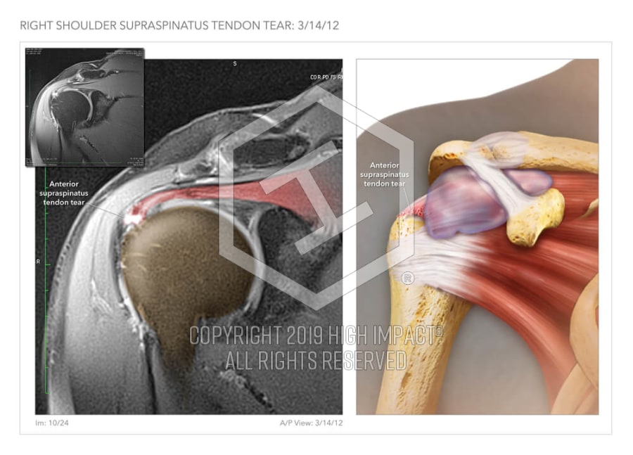
Anterior Supraspinatus Tendon Tear High Impact Visual Litigation Strategies from res.cloudinary.com The acromion can rub against (or impinge on) the tendon and the bursa, causing irritation and pain. Below are some anatomic terms doctors use to describe location (as applied to the shoulder): Tendonitis of your shoulder is an inflammation of your rotator cuff or biceps tendon. Anterior shoulder muscles, also called the pectoral muscles, attach the upper extremity to the clavicle and the thoracic cage. Shoulder muscle anatomy shoulder blade muscles body anatomy organs human body anatomy gross anatomy bicep tendonitis scapula muscular system anatomy anatomy images. A serious complication of shoulder dislocation is axillary nerve injury. Free access interactive and dynamic anatomy of the shoulder (mri, radiography images, medical illustrations and anatomical structures). 11 photos of the shoulder muscles tendons anatomy.
The subacromial bursa lies between the rotator cuff and shoulder blade and protects the tendons in this area.
Anterior shoulder dislocation an anterior dislocation accounts for 97% of recurrent or first time dislocations. The shoulder is extremely mobile and made up of several joints that work together. Axillary nerve (c5,6) from posterior cord of brachial plexus. Also in its upper part, the anterior portion of the subacromial subdeltoid bursa can be seen deep to the deltoid muscle and anterior to the biceps sheath. The muscle most commonly affected is the supraspinatus. Anterior dislocation of the shoulder joint (95% of cases) is caused by excessive extension and lateral rotation of the humerus. Deltoid tuberosity of the humerus. These muscles include the pectoralis major, pectoralis minor, subclavius and the serratus anterior muscle. Anterior fibers flex & medially rotate arm; It is the part of the shoulder that borders the chest muscles. The rotator cuff tendons can be irritated or damaged. That is there is no. The cervical spine and the biceps tendon.
The rotator cuff is a collection of muscles and tendons that surround the shoulder, giving it support and allowing a wide range of motion shoulder tendon anatomy. Deltoid tuberosity of the humerus.

0 Comments:
Posting Komentar